ECG vs. Echocardiogram – Understanding the Differences and Benefits
The prevalence of heart diseases is on the rise, posing a significant health challenge. This concerning trend is affecting individuals across various age groups and backgrounds. The factors contributing to the increase in heart diseases are complex and multifaceted, encompassing both lifestyle choices and genetic predispositions.
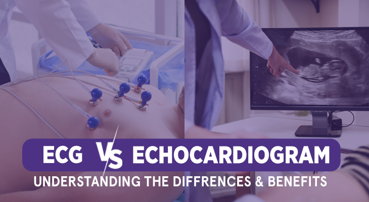
However, with technological advancement, new technologies are emerging day by day. ECGs and Echocardiograms are two such common diagnostic tests used by physicians to analyze the condition of the heart and discover disorders that affect it. Let us know more about it in detail.
Introduction to ECG and ECHO
ECG (Electrocardiogram) and ECHO (Echocardiogram) have significantly enhanced the diagnosis and management of cardiovascular conditions.
ECG, a non-invasive test, records the electrical activity of the heart and provides valuable information about its rhythm and function. This allows doctors to detect abnormalities, such as irregular heartbeats or signs of a heart attack, enabling prompt and targeted treatment.

Different types of ECG
- Resting ECG: The most common type of ECG performed. It involves attaching electrodes to specific points on the patient’s chest, limbs, and sometimes the back. The patient lies still while the ECG machine records the heart’s electrical signals at rest. This type of ECG provides valuable information about the heart’s baseline rhythm, rate, and presence of any structural abnormalities.
- Exercise Stress ECG: Also known as a stress test or treadmill test, it combines ECG monitoring with physical exercise. The patient is asked to walk on a treadmill or pedal a stationary bicycle while the ECG records the heart’s activity. This type of ECG helps evaluate the heart’s response to physical stress, identifying any abnormalities that may not be apparent at rest. It is particularly useful in diagnosing coronary artery disease and assessing exercise tolerance.
- Ambulatory ECG: This is commonly referred to as a Holter monitor, which involves wearing a portable device that continuously records the heart’s electrical activity over an extended period. This type of ECG is particularly useful in capturing intermittent abnormalities that may not be captured during a resting ECG. It allows for long-term monitoring, often spanning 24 to 48 hours or even several weeks, providing valuable insights into irregularities such as arrhythmias or symptoms like palpitations.
- Event Recorder: Event recorders are small, portable devices that patients can wear for weeks or even months. They are typically used for individuals who experience infrequent symptoms such as fainting spells or dizziness. The patient can turn on the device when they experience symptoms, which enables the recording of the heart’s electrical activity during particular events. Event recorders help correlate symptoms with any potential cardiac abnormalities, aiding in accurate diagnosis and treatment.
- Signal-Averaged ECG: This involves averaging out multiple ECG recordings to enhance the visibility of small electrical signals. It is primarily used to detect and assess the risk of ventricular arrhythmias, such as those associated with previous heart attacks or certain heart conditions.
ECHO utilizes ultrasound technology to create detailed images of the heart’s structure and function. It enables the evaluation of heart chambers, valves, and blood flow patterns, aiding in the diagnosis of conditions like heart valve abnormalities, heart failure, and congenital heart defects.

Different types of ECHO
- Transthoracic Echocardiography (TTE): This involves placing a transducer on the patient’s chest, which emits sound waves and captures the echoes as they bounce back from the heart structures. TTE provides a comprehensive evaluation of the heart’s structure, function, and blood flow patterns.
- Transesophageal Echocardiography (TEE): It involves inserting a specialized probe into the patient’s esophagus, allowing for closer proximity to the heart, and providing detailed images of the heart’s structures, valves, and blood flow. TEE is particularly useful for assessing conditions like valvular heart disease, blood clots, and infections.
- Stress Echocardiography: This combines echocardiography with physical or pharmacological stress to evaluate the heart’s function under an increased workload. During the test, images of the heart are obtained before and after exercise or medication-induced stress. Stress echocardiography helps assess coronary artery disease, exercise tolerance, and the heart’s response to stress.
- Three-Dimensional Echocardiography (3D Echo): 3D Echo creates a three-dimensional image of the heart using advanced imaging techniques. It provides a more detailed and realistic visualization of the heart’s structures, aiding in the assessment of complex cardiac conditions and assisting in surgical planning.
- Doppler Echocardiography: This measures and analyzes the blood flow through the heart and blood vessels. It helps evaluate the direction, speed, and turbulence of blood flow, providing valuable information about valve function, blood vessel abnormalities, and conditions like heart failure or congenital heart defects.
- Contrast Echocardiography: This involves the injection of a contrast agent, typically microbubbles, into a vein. These microbubbles enhance the ultrasound images, allowing better visualization of the heart’s structures and blood flow. Contrast echocardiography is useful in evaluating conditions where standard echocardiography may have limitations, such as assessing blood flow through complex structures or detecting small defects
Conclusion
Both ECG and ECHO have become indispensable tools in cardiovascular medicine, empowering physicians to make accurate diagnoses and develop personalized treatment plans for patients, ultimately improving their cardiac health outcomes.
Accord Hospital in Faridabad, Delhi NCR, is renowned for its exceptional cardiac care hospital and is home to some of the best cardiologists in the region. With a team of highly skilled and experienced heart doctors, Accord Hospital is committed to providing comprehensive and personalized care for patients with cardiovascular conditions. Equipped with state-of-the-art facilities and cutting-edge technology, the hospital offers advanced diagnostic services, making it the go-to heart diagnostic centre in Faridabad. Patients can trust Accord Hospital for accurate diagnoses, effective treatment plans, and compassionate care, ensuring the best possible outcomes for their heart health.